

- Quadre mèdic
- Especialitats
- Unitats especialitzades
 Institut del Cor Teknon
 Unitat d’Obesitat
 Institut Oncològic
 Unitat d’Medicina i cirurgia sense sang
 Institut de Neurociències
 Unitat d’atenció al lesionat de trànsit
 Unitat de Pneumologia
 Unitat de Medicina Marítima
 Institut de Teràpia Regenerativa Tissular
 Unitat de Tractament del Dolor
 Clínica del Tennis
 Unitat de malalties inflamatòries i autoimmunes sistèmiques
 Unitat de Reproducció Assistida
 Unitat del Son
 Unitat de Síndromes de Sensibilització Central
 Unitat del Virus del Papil·loma Humà
- Unitats especialitzades
- Àrea diagnòstica
- Proves diagnòstiques
 Diagnòstic per la ImatgeExploracions diagnòstiques i intervencionistes.
 Laboratori d’anatomia patològicaDisposa d’una segona opinió per part d’especialistes.
 Laboratori d’Anàlisis ClíniquesServei integral a l’àrea clínica.
 EndoscòpiaDiagnòstic precís sense utilitzar la cirurgia.
 ElectrofisiologiaExploració funcional del Sistema Nerviós Central.
 ElectromiografíaAvaluació clínica i neurofisiològica de la patologia neuromuscular.
 DensitometriaTècnica diagnòstica per comprovar la densitat mineral de l’os.
 UrodinàmiaDiagnòstic dels trastorns de micció i incontinència.
- Revisions mèdiques

 GeneralUn control intel·ligent de la teva salut.
 CompletUn examen exhaustiu de la teva salut.
 Complet PlusLa nostra revisió mèdica més exhaustiva.
 ViatgersSi vols fer un viatge, la teva salut forma part de l’equipatge.
 EsportiuUna revisió a fons per a potenciar el teu rendiment.
 CardiològicUna bona notícia és saber que el teu cor està sota control.
 Per a empresesUna eina que potencia la satisfacció, productivitat i fidelització del treballador.
- Proves diagnòstiques
- El nostre centre
- Entorn assistencial
 HospitalitzacióAmb habitacions lluminoses, funcionals i completament equipades.
 Unitat de SemicriticsDotada de tecnologia per al diagnòstic i tractament que requereixen una vigilància i cura especial.
 Programa d’Alimentació SaludableVolem millorar la salut de les persones, per això promovem una alimentació saludable, conscient i sostenible als nostres hospitals.
 InfermeriaUn equip de més de 400 professionals.
 Servei d’Urgències24 hores al dia al teu servei sense interrupcions.
 Àrea Pacient PrivatBasat en l’alta qualitat i el servei personalitzat, oferim un conjunt de serveis que complementen les cures mèdico-assistencials
 L’Àrea QuirúrgicaDisposa de 20 quiròfans, 12 d’ells preparats per a la cirurgia d’alt risc.
 International programUn assistent d’aquest programa us oferirà atenció integral i personalitzada
 Comitè d’Ètica AssistencialAjuda els ciutadans i als professionals de la salut a orienta la seva actuació en casos de conflictes morals.
 UCI-UCUnitat polivalent que disposa de boxes completament equipats amb el sistema més modern de monitorització.
 Atenció al PacientA disposició de tots els pacients i acompanyants del centre.
 InvestigacióLa investigació constitueix un dels pilars bàsics de Centre Mèdic Teknon.
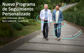 Programa de Seguiment PersonalitzatT’acompanyem durant el teu procés mèdic. Organitzem i agendem les teves cites i proves.
 Qualitat i Seguretat del PacientAdoptem models de gestió basats en els estàndards més exigents nacionals i internacionals.
- Entorn assistencial
- Actualitat
- Actualitat
 NotíciesConeix el que està passant a Centro Médico Teknon. Consulta la nostra secció de notícies.
 AgendaPots trobar tots els esdeveniments que hem organitzat sobre salut i aquells temes d’actualitat que et poden interessar. Accedeix a la nostra agenda d’activitats.
 VídeosEn aquesta secció trobaràs una àmplia col·lecció de vídeos relacionats amb les nostres especialitats.
 PodcastTemes mèdics d’actualitat, tractaments innovadors, consells de salut y experiències de pacients abordabats pels nostres especialistes.
 Continguts de salut
- Actualitat
- Blog

- Quadre mèdic
- Especialitats
- Unitats especialitzades
- Institut del Cor Teknon
- Unitat d’Obesitat
- Institut Oncològic
- Unitat d’Medicina i cirurgia sense sang
- Institut de Neurociències
- Unitat d’atenció al lesionat de trànsit
- Unitat de Pneumologia
- Unitat de Medicina Marítima
- Institut de Teràpia Regenerativa Tissular
- Unitat de Tractament del Dolor
- Clínica del Tennis
- Unitat de malalties inflamatòries i autoimmunes sistèmiques
- Unitat de Reproducció Assistida
- Unitat del Son
- Unitat de Síndromes de Sensibilització Central
- Unitat del Virus del Papil·loma Humà
- Unitats especialitzades
- Àrea diagnòstica
- Proves diagnòstiques
- Diagnòstic per la ImatgeExploracions diagnòstiques i intervencionistes.
- Laboratori d’anatomia patològicaDisposa d’una segona opinió per part d’especialistes.
- Laboratori d’Anàlisis ClíniquesServei integral a l’àrea clínica.
- EndoscòpiaDiagnòstic precís sense utilitzar la cirurgia.
- ElectrofisiologiaExploració funcional del Sistema Nerviós Central.
- ElectromiografíaAvaluació clínica i neurofisiològica de la patologia neuromuscular.
- DensitometriaTècnica diagnòstica per comprovar la densitat mineral de l’os.
- UrodinàmiaDiagnòstic dels trastorns de micció i incontinència.
- Revisions mèdiques
- GeneralUn control intel·ligent de la teva salut.
- CompletUn examen exhaustiu de la teva salut.
- Complet PlusLa nostra revisió mèdica més exhaustiva.
- ViatgersSi vols fer un viatge, la teva salut forma part de l’equipatge.
- EsportiuUna revisió a fons per a potenciar el teu rendiment.
- CardiològicUna bona notícia és saber que el teu cor està sota control.
- Per a empresesUna eina que potencia la satisfacció, productivitat i fidelització del treballador.
- Proves diagnòstiques
- El nostre centre
- Entorn assistencial
- HospitalitzacióAmb habitacions lluminoses, funcionals i completament equipades.
- Unitat de SemicriticsDotada de tecnologia per al diagnòstic i tractament que requereixen una vigilància i cura especial.
- Programa d’Alimentació SaludableVolem millorar la salut de les persones, per això promovem una alimentació saludable, conscient i sostenible als nostres hospitals.
- InfermeriaUn equip de més de 400 professionals.
- Servei d’Urgències24 hores al dia al teu servei sense interrupcions.
- Àrea Pacient PrivatBasat en l’alta qualitat i el servei personalitzat, oferim un conjunt de serveis que complementen les cures mèdico-assistencials
- L’Àrea QuirúrgicaDisposa de 20 quiròfans, 12 d’ells preparats per a la cirurgia d’alt risc.
- International programUn assistent d’aquest programa us oferirà atenció integral i personalitzada
- Comitè d’Ètica AssistencialAjuda els ciutadans i als professionals de la salut a orienta la seva actuació en casos de conflictes morals.
- UCI-UCUnitat polivalent que disposa de boxes completament equipats amb el sistema més modern de monitorització.
- Atenció al PacientA disposició de tots els pacients i acompanyants del centre.
- InvestigacióLa investigació constitueix un dels pilars bàsics de Centre Mèdic Teknon.
- Programa de Seguiment PersonalitzatT’acompanyem durant el teu procés mèdic. Organitzem i agendem les teves cites i proves.
- Qualitat i Seguretat del PacientAdoptem models de gestió basats en els estàndards més exigents nacionals i internacionals.
- Entorn assistencial
- Actualitat
- Actualitat
- NotíciesConeix el que està passant a Centro Médico Teknon. Consulta la nostra secció de notícies.
- AgendaPots trobar tots els esdeveniments que hem organitzat sobre salut i aquells temes d’actualitat que et poden interessar. Accedeix a la nostra agenda d’activitats.
- VídeosEn aquesta secció trobaràs una àmplia col·lecció de vídeos relacionats amb les nostres especialitats.
- PodcastTemes mèdics d’actualitat, tractaments innovadors, consells de salut y experiències de pacients abordabats pels nostres especialistes.
- Continguts de salut
- Actualitat
- Blog


- Especialitats
 Fernández Agrafojo Dora
- Els nostres serveis
- Retina (DMAE)
 Centro Médico Teknon
 Centro Médico Teknon

La retina, la capa més interna del globus ocular, és una membrana transparent formada per nombroses cèl·lules fotosensibles encarregades de rebre els estímuls lluminosos i transmetre'ls a través dels seus terminals nerviosos al cervell. Existeixen dos tipus de cèl·lules fotoreceptores: els cons i els bastons. Els cons funcionen millor amb llum diürna i estan especialitzats en la visió dels colors. Els bastons són més nombrosos i funcionen amb llum nocturna o a la foscor. Els cons són més abundants al centre de la retina, també anomenada màcula o fòvea, i els bastons a la perifèria de la mateixa. Quan cons i bastons són estimulats per la llum, es generen impulsos que són transmesos a través de les seves fibres nervioses i conflueixen per formar el nervi òptic.
La retina es nodreix per vasos arterials retinians i per capilars de la coroides, que és una capa vascularitzada situada per fora de la retina.
- The vitreous
The vitreous is a transparent, gelatinous substance which occupies the inner part of the eye, providing support to the retina. It has no nutritive vessels, since it would lose its transparency. Over the years, the vitreous may lose its consistency and separate from the retina, causing deposits which are perceived as threads or spots that move with the eyes; these are more visible in bright light or when looking at light-colored walls or objects. These deposits are referred to as myodesopsia, "floaters", or "mouches volantes". Usually they do not indicate any kind of disease, nor do they represent any risk to one's vision; however, if they are perceived, one must rule out the presence of any coexisting pathology in the retina or vitreous.
- What are the main problems of the retina?
The retina can be affected by:
- Many general diseases, such as diabetes or ocular hypertension (high eye pressure)
- Inflammations or infections
- Vascular irregularities such as thrombosis or embolisms
- detachments which produce loss of vision in one part of the field of vision, or in its entirety
- Degeneration, its most frequent and important instance being age-related macular degeneration
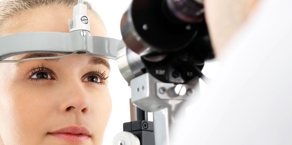
- How to diagnose these diseases?
Diagnosis of all these diseases is important in order to provide early treatment. For this purpose we have many techniques and instruments at our disposal, most notably: retinoscopy (observation of the retina and vitreous with or without dilating the pupil), fluorescein angiography, computerized campimetry, and for some time now, optical coherence tomography – a very high-precision, latest-generation instrument for studying the retina.
Irregularities in the layers of the retina which nourish and provide oxygen to the pigment epithelium are the cause of age-related macular degeneration (AMD). This can be either dry or wet (exudative) AMD, the latter being characterized by the appearance of blood vessels, which can give rise to hemorrhaging and accumulation of liquid between layers of the retina.
With AMD, the patient will report irregularity in the central vision of the affected eye, image distortion or deformity.In such cases, the Amsler test offers a simple method for monitoring symptoms.
Recent years have brought to light the so-called antiangiogenic substances. When injected into the inner eye, they often make it possible to control loss of vision or even to improve it, in some cases of exudative (wet) age-related macular degeneration.
With the help of the latest technology in ophthalmological exploration, we are able to detect retinopathies in the earliest stages of macular degeneration, when the patient may not report any kind of symptoms. This is the case with Optical Coherence Tomography (OCT).

This technique examines retinal layers through images, visualizing a cross-section of the desired area of the retina. The slightest irregularity among the layers (as in cases of AMD) is projected, measured, compared and monitored, thanks to the different visual perspectives offered by the software. Even 3-D imaging of the retina can be obtained.
A database of population studies is incorporated, giving us automatic comparisons of the thickness of retinal ganglion fibers which lead to the optic nerve, and in this way assess their normality. This is especially useful in the case of patients with glaucoma, both as a support to diagnosis as well as in monitoring its development over time.