

- Cuadro médico
- Especialidades
- Unidades especializadas
 Instituto del Corazón
 Unidad de Obesidad
 Instituto Oncológico
 Unidad de Medicina y Cirugía sin sangre
 Instituto de Neurociencias
 Unidad de Atención al Lesionado de Tráfico
 Instituto de Neumología
 Unidad de Medicina Marítima
 Instituto de Terapia Regenerativa Tisular
 Unidad de Tratamiento del Dolor
 Clínica del Tenis
 Unidad de Enfermedades Inflamatorias y Autoinmunes
 Unidad de Reproducción
Asistida
Unidad de Reproducción
Asistida Unidad del Sueño
 Unidad de Síndromes de Sensibilización Central
 Unidad del Virus del Papiloma Humano
- Unidades especializadas
- Área diagnóstica
- Pruebas diagnósticas
 Diagnóstico por la imagenExploraciones diagnósticas e intervencionistas.
 Laboratorio de anatomía patológicaDispone de una segunda opinión de la mano de especialistas.
 Laboratorio de análisis clínicosServicio integral en el área clínica.
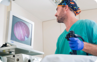 EndoscopiaDiagnóstico preciso sin utilizar la cirugía.
 ElectrofisiologíaExploración funcional del Sistema Nervioso Central.
 ElectromiografíaEvaluación clínica y neurofisiológica de la patología neuromuscular.
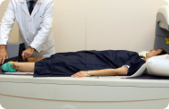 DensitometríaTécnica diagnóstica para comprobar la densidad mineral del hueso.
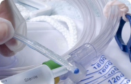 UrodinamiaDiagnóstico de los trastornos de micción e incontinencia.
- Chequeos médicos

 GeneralUn control inteligente de tu salud
 CompletoUn examen exhaustivo de tu salud
 Completo PlusNuestro chequeo más exclusivo
 ViajerosSi vas a emprender un viaje, tu salud es parte del equipaje
 DeportivoUna revisión a fondo para potenciar tu rendimiento
 CardiológicoUna buena noticia es saber que tu corazón está bajo control
 Para empresasUna herramienta que potencia la satisfacción, productividad y fidelización del empleado
- Pruebas diagnósticas
- Nuestro Centro
- Entorno asistencial Teknon
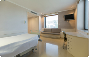 HospitalizaciónHabitaciones luminosas, funcionales y completamente equipadas.
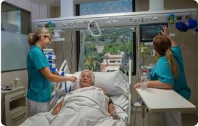 Unidad de SemicríticosDotada de tecnología para diagnósticos y tratamientos que requieren cuidado especial.
 Programa de Alimentación SaludableQueremos mejorar la salud de las personas, por ello promovemos una alimentación saludable, consciente y sostenible en nuestros hospitales.
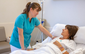 EnfermeríaEquipo de más de 400 profesionales.
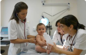 Servicio de Urgencias24 horas al día a tu servicio sin interrupciones.
 Área Paciente PrivadoBasado en la alta calidad y el servicio personalizado, ofrecemos un conjunto de servicios que complementan los cuidados médico-asistenciales
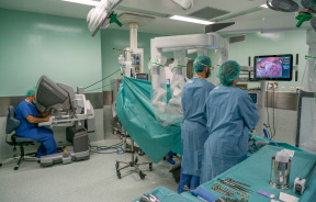 Área QuirúrgicaCuenta con 20 quirófanos, 12 de ellos preparados para cirugía de alto riesgo.
 Programa internacionalUn asistente de este programa le ofrecerá atención integral y personalizada
 Comité de Ética AsistencialAyuda a ciudadanos y profesionales a orientar su actuación en casos de conflictos morales.
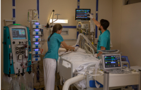 UCI-UCUnidad polivalente con boxes equipados con modernos sistemas de monitorización.
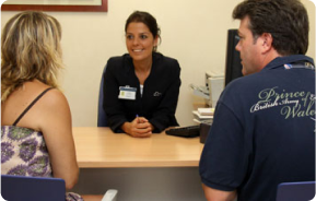 Atención al pacienteA disposición de todos los pacientes y acompañantes del centro.
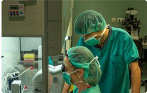 InvestigaciónLa investigación constituye uno de los pilares básicos de Centro Médico Teknon.
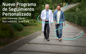 Programa de Seguimiento PersonalizadoTe acompañamos durante tu proceso médico. Organizamos y agendamos tus citas y pruebas.
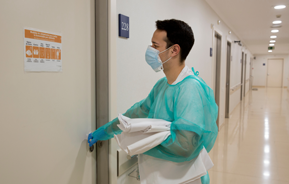 Calidad y Seguridad del PacienteAdoptamos modelos de gestión basados en los estándares más exigentes nacionales e internacionales.
- Entorno asistencial Teknon
- Actualidad
- Actualidad
 NoticiasConoce qué está pasando en Centro Médico Teknon. Consulta nuestra sección de noticias.
 AgendaPuedes encontrar todos los eventos que hemos organizado sobre salud y aquellos temas de actualidad que te pueden interesar. Accede a nuestra agenda de actividades.
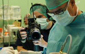 VídeosEn esta sección encontrarás una amplia colección de videos relacionados con nuestras especialidades.
 PodcastTemas médicos de actualidad, tratamientos innovadores, consejos de salud y experiencias de pacientes abordados por nuestros especialistas.
 Contenidos de salud
- Actualidad
- Blog

- Cuadro médico
- Especialidades
- Unidades especializadas
- Instituto del Corazón
- Unidad de Obesidad
- Instituto Oncológico
- Unidad de Medicina y Cirugía sin sangre
- Instituto de Neurociencias
- Unidad de Atención al Lesionado de Tráfico
- Instituto de Neumología
- Unidad de Medicina Marítima
- Instituto de Terapia Regenerativa Tisular
- Unidad de Tratamiento del Dolor
- Clínica del Tenis
- Unidad de Enfermedades Inflamatorias y Autoinmunes
- Unidad de Reproducción
Asistida
- Unidad del Sueño
- Unidad de Síndromes de Sensibilización Central
- Unidad del Virus del Papiloma Humano
- Unidades especializadas
- Área diagnóstica
- Pruebas diagnósticas
- Diagnóstico por la imagenExploraciones diagnósticas e intervencionistas.
- Laboratorio de anatomía patológicaDispone de una segunda opinión de la mano de especialistas.
- Laboratorio de análisis clínicosServicio integral en el área clínica.
- EndoscopiaDiagnóstico preciso sin utilizar la cirugía.
- ElectrofisiologíaExploración funcional del Sistema Nervioso Central.
- ElectromiografíaEvaluación clínica y neurofisiológica de la patología neuromuscular.
- DensitometríaTécnica diagnóstica para comprobar la densidad mineral del hueso.
- UrodinamiaDiagnóstico de los trastornos de micción e incontinencia.
- Chequeos médicos
- GeneralUn control inteligente de tu salud
- CompletoUn examen exhaustivo de tu salud
- Completo PlusNuestro chequeo más exclusivo
- ViajerosSi vas a emprender un viaje, tu salud es parte del equipaje
- DeportivoUna revisión a fondo para potenciar tu rendimiento
- CardiológicoUna buena noticia es saber que tu corazón está bajo control
- Para empresasUna herramienta que potencia la satisfacción, productividad y fidelización del empleado
- Pruebas diagnósticas
- Nuestro Centro
- Entorno asistencial Teknon
- HospitalizaciónHabitaciones luminosas, funcionales y completamente equipadas.
- Unidad de SemicríticosDotada de tecnología para diagnósticos y tratamientos que requieren cuidado especial.
- Programa de Alimentación SaludableQueremos mejorar la salud de las personas, por ello promovemos una alimentación saludable, consciente y sostenible en nuestros hospitales.
- EnfermeríaEquipo de más de 400 profesionales.
- Servicio de Urgencias24 horas al día a tu servicio sin interrupciones.
- Área Paciente PrivadoBasado en la alta calidad y el servicio personalizado, ofrecemos un conjunto de servicios que complementan los cuidados médico-asistenciales
- Área QuirúrgicaCuenta con 20 quirófanos, 12 de ellos preparados para cirugía de alto riesgo.
- Programa internacionalUn asistente de este programa le ofrecerá atención integral y personalizada
- Comité de Ética AsistencialAyuda a ciudadanos y profesionales a orientar su actuación en casos de conflictos morales.
- UCI-UCUnidad polivalente con boxes equipados con modernos sistemas de monitorización.
- Atención al pacienteA disposición de todos los pacientes y acompañantes del centro.
- InvestigaciónLa investigación constituye uno de los pilares básicos de Centro Médico Teknon.
- Programa de Seguimiento PersonalizadoTe acompañamos durante tu proceso médico. Organizamos y agendamos tus citas y pruebas.
- Calidad y Seguridad del PacienteAdoptamos modelos de gestión basados en los estándares más exigentes nacionales e internacionales.
- Entorno asistencial Teknon
- Actualidad
- Actualidad
- NoticiasConoce qué está pasando en Centro Médico Teknon. Consulta nuestra sección de noticias.
- AgendaPuedes encontrar todos los eventos que hemos organizado sobre salud y aquellos temas de actualidad que te pueden interesar. Accede a nuestra agenda de actividades.
- VídeosEn esta sección encontrarás una amplia colección de videos relacionados con nuestras especialidades.
- PodcastTemas médicos de actualidad, tratamientos innovadores, consejos de salud y experiencias de pacientes abordados por nuestros especialistas.
- Contenidos de salud
- Actualidad
- Blog


- Especialidades
 Fernández Agrafojo Dora
- Servicios
- Retina (DMAE)

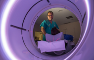
 Centro Médico Teknon
 Centro Médico Teknon

La retina, la capa más interna del globo ocular,es una membrana transparente compuesta de numerosas células fotosensibles encargadas de recibir los estímulos luminosos y transmitirlos a través de sus terminales nerviosas al cerebro. Existen 2 tipos de células fotoreceptoras: los conos y los bastones.
Los conos funcionan mejor con luz diurna y están especializados en la visión de los colores. Los bastones son más numerosos y funcionan con la luz nocturna o la oscuridad. Los conos son más abundantes en el centro de la retina, llamada también mácula o fóvea. Los bastones se encuentran en la periferia. Cuando conos y bastones son estimulados por la luz, se generan impulsos que son transmitidos a través de las fibras nerviosas de los mismos y que confluyen para formar el nervio óptico.
La retina se nutre por vasos arteriales retinianos y por capilares de la coroides, que es la capa vascularizada más externa de la retina.
- ¿Qué es el vítreo?
El vítreo es una sustancia transparente, de consistencia gelatinosa, que ocupa el interior del ojo, proporcionando apoyo a la retina. No tiene vasos nutricios, pues perdería su transparencia. Con los años el vítreo pierde su consistencia y se separa de la retina, dando lugar a condensaciones, que se perciben como hilos o manchas que se mueven con el movimiento de los ojos y son más visibles en condiciones de mucha luz o al mirar paredes u objetos claros. Esas condensaciones reciben el nombre de miodesopsias o "moscas volantes", y usualmente no son signo de enfermedad alguna y no representan ningún riesgo para la visión. Sin embargo, al percibirlas, es necesario descartar la presencia de alguna patología coexistente en la retina o vítreo.
- ¿Cuáles son los principales problemas de la retina?
La retina puede verse afectada por:
- Múltiples enfermedades generales, como por ejemplo diabetes o hipertensión ocular
- Inflamaciones o infecciones
- Alteraciones vasculares como trombosis o embolias
- Desprendimientos que pueden manifestarse por la pérdida de la visión en una parte del campo visual o en su totalidad
- Degeneraciones, siendo en este último caso la más frecuente e importante la degeneración macular asociada a la edad (DMAE)
- ¿Cómo se diagnostican estas enfermedades?
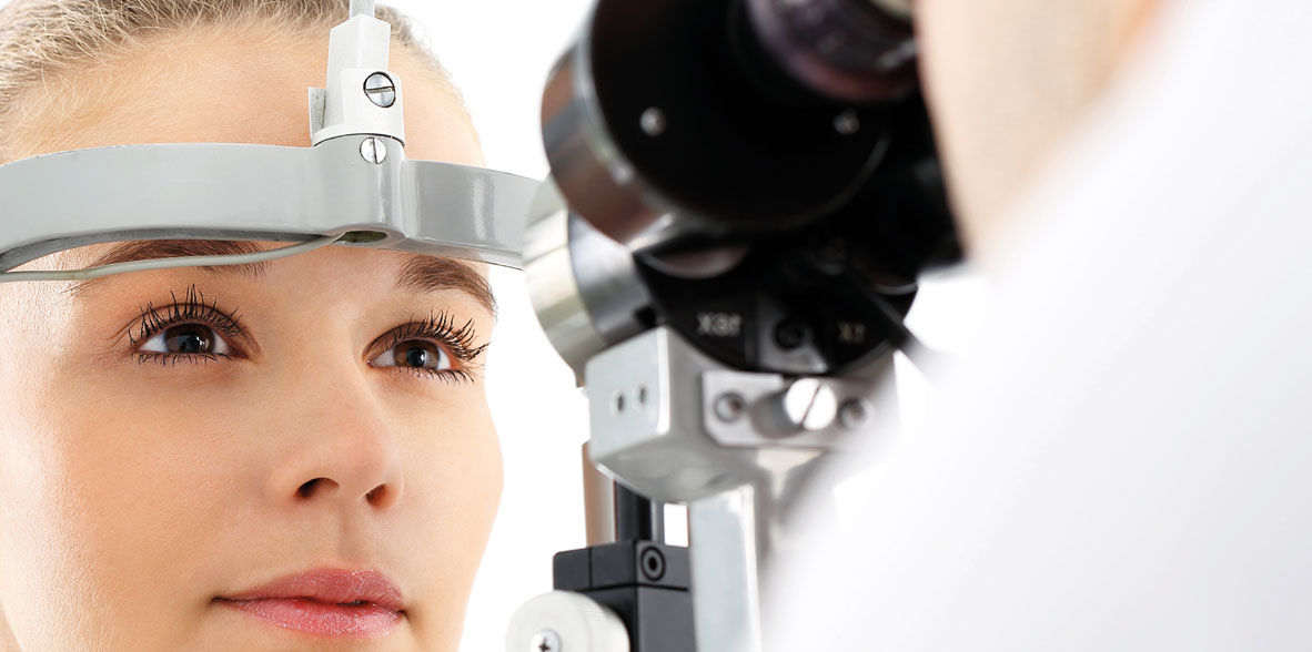
Diagnosticar todas estas enfermedades es importante para su pronto tratamiento, y para ello contamos con numerosas técnicas y aparatos, destacando entre ellas la retinoscopía (u observacion de la retina y vítreo con dilatación o no de la pupila), la angiografía fluoresceínica, la campimetría computerizada y desde hace algún tiempo con un instrumento de altísima precisión en el estudio de la retina como es el tomógrafo de coherencia óptica (OCT) de última generación.
La alteración en las capas de la retina, asociadas al epitelio pigmentario y encargadas de nutrirla y oxigenarla, son la causa de la aparición de la degeneración macular asociada a la edad (DMAE). Ésta puede ser de tipo seca o húmeda (exudativa), caracterizándose por la aparición de vasos sanguíneos en el caso exudativo, pudiendo dar lugar a hemorragias y acúmulo de líquido entre las capas de la retina.
Frente a la DMAE el paciente referirá alteración en la visión central del ojo afectado, distorsión y deformidad de la imagen. El test de Amsler permitirá en esos casos monitorizar la sintomatología de modo sencillo.
En estos últimos años han aparecido sustancias llamadas antiangiogénicas, que inyectadas en el interior del ojo nos permiten en muchos casos controlar la pérdida de visión e incluso en ocasiones mejorarla en casos de degeneración macular asociada a la edad de tipo exudativo (húmeda).
Contar con los últimos avances técnicos dentro de la exloración oftalmológica en casos de degeneración macular, nos permitirá detectar las retinopatías incluso en fases muy incipientes, donde el paciente puede no referir aún sintomatología alguna. Este es el caso de la Tomografía de coherencia óptica (OCT).
 La tomografía de coherencia óptica (OCT) es una técnica que evalúa mediante imágenes las capas de la retina, visualizándose un corte transversal de la zona de la retina deseada. Ante la más mínima alteración entre capas (como en los casos de DMAE) y gracias a las diferentes prespectivas visuales que ofrece su software, cualquier alteración quedará proyectada, medida, comparada y monitorizada. Permite incluso la obtención en 3D de la retina evaluada.
Cuenta con una base de datos (estudios de población), que compara automáticamente el espesor de las fibras ganglionares del la retina que se dirigen al nervio óptico para valorar su rango de normalidad. Muy útil en el caso de pacientes con glaucoma, tanto como apoyo en diagnóstico como en su seguimiento en futuras revisiones.