




- Unitats mèdiques
- Especialitats
- Consultoris
- Quadre Mèdic
- Àrea diagnòstica
- El nostre centre
- Actualitat


- Unitats mèdiques
- Especialitats
- Consultoris
- Quadre Mèdic
- Àrea diagnòstica
- El nostre centre
- Actualitat


- Àrea diagnòstica
 Proves diagnòstiques
- Tractaments i especialitats
- Diagnòstic per la Imatge
- Ressonància Magnètica
Ressonància Magnètica
La Ressonància Magnètica és una prova diagnòstica no invasiva (i que no utilitza raigs X) que consisteix en l’obtenció d’imatges d’alta definició anatòmica en base a la ressonància que es produeix en els teixits sotmesos a un camp magnètic. És la tècnica d’imatge que proporciona una major diferenciació entre els diferents teixits de l’organisme, i que permet obtenir informació molt precisa sobre l’estructura i la composició de l’òrgan que s’ha d’analitzar.
- Hospital Quirón Teknon compta amb la primera Ressonància Magnètica Oberta d'Alt Camp a Catalunya. Així mateix, també permet realitzar l'exploració, no solament amb el pacient estirat, sinó amb les articulacions flexionades i en moviment.
- Neurorradiología
- RM Cerebral (craneal)
 Prueba diagnóstica no invasiva que consiste en la obtención de imágenes de alta definición anatómica del cerebro mediante el empleo de un campo electromagnético y ondas de radio (con un emisor y un receptor). No utiliza radiación ionizante. Indicaciones: problemas vasculares, pérdida de memoria, epilepsia, cefalea, malformaciones, sospecha de tumor, meningitis.
 - RM Cais
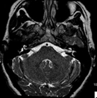 Prueba diagnóstica no invasiva que consiste en la obtención de imágenes de alta definición anatómica del oído interno mediante el empleo de un campo electromagnético y ondas de radio (con un emisor y un receptor). No utiliza radiación ionizante. Indicaciones: problemas auditivos, mareos, vértigos.
 - RM Cuello
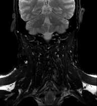 Prueba diagnóstica no invasiva que consiste en la obtención de imágenes de alta definición anatómica del cuello mediante el empleo de un campo electromagnético y ondas de radio (con un emisor y un receptor). No utiliza radiación ionizante. Indicaciones: sospecha de tumor, infecciones, ganglios.
 - Angio-RM intracraneal
 Prueba diagnóstica no invasiva que consiste en la obtención de imágenes de alta definición anatómica de las arterias cerebrales mediante el empleo de un campo electromagnético y ondas de radio (con un emisor y un receptor). No utiliza radiación ionizante. En la mayoría de los casos es necesario el empleo de contraste paramagnético (Gadolinio). Permite un estudio angiográfico no invasivo gracias a la inyección de Gadolinio con posterior reconstrucción en 2D y 3D, gracias a estaciones de trabajo especializadas. Indicaciones: malformaciones vasculares, aneurismas de arterias cerebrales, arteriosclerosis.
 - Angio-RM troncos supraaórticos
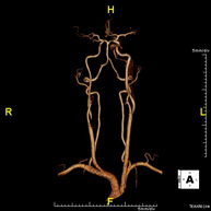 Prueba diagnóstica no invasiva que consiste en la obtención de imágenes de alta definición anatómica de las arterias carótidas y vertebrales a nivel de cuello, mediante el empleo de un campo electromagnético y ondas de radio (con un emisor y un receptor). No utiliza radiación ionizante. En la mayoría de los casos es necesario el empleo de contraste paramagnético (Gadolinio). Permite un estudio angiográfico no invasivo gracias a la inyección de Gadolinio con posterior reconstrucción en 2D y 3D, gracias a estaciones de trabajo especializadas. Indicaciones: problemas circulatorios cerebrales, síncope.
 - RM Espectroscopia Cerebral
Prueba diagnóstica no invasiva que consiste en la obtención de imágenes de alta definición anatómica del cerebro mediante el empleo de un campo electromagnético y ondas de radio (con un emisor y un receptor). No utiliza radiación ionizante. Se realiza, posteriormente, una medición cualitativa y cuantitativa de varios metabolitos (sustancias), que permitirán caracterizar las lesiones. Indicaciones: tumores, trastornos metabólicos, infecciones, epilepsia.
- RM Estudio Flujo LCR
Prueba diagnóstica no invasiva que consiste en la obtención de imágenes de alta definición anatómica del sistema ventricular cerebral, mediante el empleo de un campo electromagnético y ondas de radio (con un emisor y un receptor). No utiliza radiación ionizante. Indicaciones: estudio de hidrocefalia y estenosis del acueducto encefálico.
- RM Columna cervical
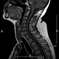 Prueba diagnóstica no invasiva que consiste en la obtención de imágenes de alta definición anatómica de la columna cervical mediante el empleo de un campo electromagnético y ondas de radio (con un emisor y un receptor). No utiliza radiación ionizante. Indicaciones: traumatismo, degeneración de la columna, hernias.
 - RM Columna dorsal
 Prueba diagnóstica no invasiva que consiste en la obtención de imágenes de alta definición anatómica de la columna dorsal, mediante el empleo de un campo electromagnético y ondas de radio (con un emisor y un receptor). No utiliza radiación ionizante. Indicaciones: traumatismo, problemas degenerativos, hernias, tumores.
 - RM Columna lumbar
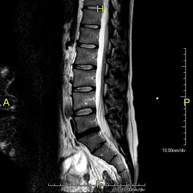 Prueba diagnóstica no invasiva que consiste en la obtención de imágenes de alta definición anatómica de la lumbar y sacra, mediante el empleo de un campo electromagnético y ondas de radio (con un emisor y un receptor). No utiliza radiación ionizante. Indicaciones: traumatismos, ciática, hernias discales, tumores, infecciones.
 - RM Mielografía
 Prueba diagnóstica no invasiva que consiste en la obtención de imágenes de alta definición anatómica de la médula espinal y las raíces nerviosas mediante el empleo de un campo electromagnético y ondas de radio (con un emisor y un receptor), así como secuencias de estudio especiales (mielográficas). No utiliza radiación ionizante. Indicaciones: hernias, compresiones medulares.
 - RM Plexo braquial
 Prueba diagnóstica no invasiva que consiste en la obtención de imágenes de alta definición anatómica de los nervios que conforman el plexo del brazo mediante el empleo de un campo electromagnético y ondas de radio (con un emisor y un receptor). No utiliza radiación ionizante. Indicaciones: estudio de parálisis de extremidades superiores para planificación quirúrgica, lesiones tumorales.
 - RM Columna cervical + dorsal
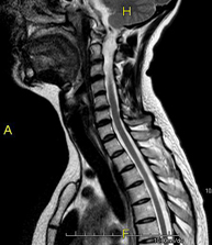 Prueba diagnóstica no invasiva que consiste en la obtención de imágenes de alta definición anatómica de la columna cervical y dorsal mediante el empleo de un campo electromagnético y ondas de radio (con un emisor y un receptor). No utiliza radiación ionizante. Indicaciones: traumatismo, degeneración de la columna, hernias.
 - RM Columna dorsal + lumbar
 Prueba diagnóstica no invasiva que consiste en la obtención de imágenes de alta definición anatómica de la columna dorsal y lumbar, mediante el empleo de un campo electromagnético y ondas de radio (con un emisor y un receptor). No utiliza radiación ionizante. Indicaciones: traumatismo, degeneración de la columna, hernias.
 - RM Columna completa
Prueba diagnóstica no invasiva que consiste en la obtención de imágenes de alta definición anatómica de la columna cervical, dorsal y lumbar, mediante el empleo de un campo electromagnético y ondas de radio (con un emisor y un receptor). No utiliza radiación ionizante. Indicaciones: detección de metástasis, escoliosis, traumatismo.
- RM Sacroilíacas
 Prueba diagnóstica no invasiva que consiste en la obtención de imágenes de alta definición anatómica de las articulaciones sacroilíacas mediante el empleo de un campo electromagnético y ondas de radio (con un emisor y un receptor). No utiliza radiación ionizante. Indicaciones: dolor sacroilíaco inflamatorio.
 - RM ATM (Articulación Temporo-Mandibular)
 Prueba diagnóstica no invasiva que consiste en la obtención de imágenes de alta definición anatómica de las articulaciones témporo-mandibulares mediante el empleo de un campo electromagnético y ondas de radio (con un emisor y un receptor). No utiliza radiación ionizante. Se realiza un estudio funcional de las ATM, en las distintas fases de apertura y cierre de la boca. Indicaciones: dolor, bloqueo de la articulación y ruidos durante la masticación.
 - RM Órbitas
 Prueba diagnóstica no invasiva que consiste en la obtención de imágenes de alta definición anatómica de las órbitas mediante el empleo de un campo electromagnético y ondas de radio (con un emisor y un receptor). No utiliza radiación ionizante. Indicaciones: visión doble, traumatismo, sospecha de tumor, hipertiroidismo.
 - RM Hipófisis
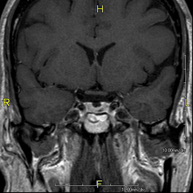 Prueba diagnóstica no invasiva que consiste en la obtención de imágenes de alta definición anatómica de la hipófisis cerebral mediante el empleo de un campo electromagnético y ondas de radio (con un emisor y un receptor). No utiliza radiación ionizante. Indicaciones: trastorno del crecimiento, trastornos hormonales, tumores.
 - RM Senos paranasales
 Prueba diagnóstica no invasiva que consiste en la obtención de imágenes de alta definición anatómica de los senos paranasales mediante el empleo de un campo electromagnético y ondas de radio (con un emisor y un receptor). No utiliza radiación ionizante. Indicaciones: estudio de tumores.
- Tórax
- RM Mediastino
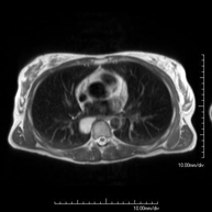 Prueba diagnóstica no invasiva que consiste en la obtención de imágenes de alta definición anatómica del mediastino mediante el empleo de un campo electromagnético y ondas de radio (con un emisor y un receptor). No utiliza radiación ionizante. El mediastino es la parte central de la caja torácica que incluye el timo, los grandes vasos (aorta torácica, vena cava inferior y superior, etc.), el corazón, la tráquea y los bronquios principales, ganglios linfáticos mediastínicos e hiliares, el esófago, etc. Está especialmente indicada en lesiones mediastínicas para diferenciar si son quísticas o sólidas, en el diagnóstico diferencial de las lesiones del mediastino anterior, etc. En ocasiones se deberá emplear contraste paramagnético (Gadolinio) para completar el estudio.
 - RM Tórax
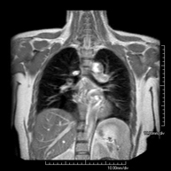 Prueba diagnóstica no invasiva que consiste en la obtención de imágenes de alta definición anatómica del tórax mediante el empleo de un campo electromagnético y ondas de radio (con un emisor y un receptor). No utiliza radiación ionizante. Está indicada en aquellas lesiones pulmonares en las que debe descartarse si existe infiltración del mediastino o de la pared torácica, para diferenciar si una lesión torácica es sólida o quística, etc. En ocasiones se deberá emplear contraste paramagnético (Gadolinio) para completar el estudio.
 - RM Pared Torácica
 Prueba diagnóstica no invasiva que consiste en la obtención de imágenes de alta definición anatómica de la pared torácica mediante el empleo de un campo electromagnético y ondas de radio (con un emisor y un receptor). No utiliza radiación ionizante. Está indicada en el estudio de lesiones de pared torácica: costales, esternales, musculares (pectorales, intercostales, paravertebrales, etc.), en el estudio de dolores costales, etc. En ocasiones se deberá emplear contraste paramagnético (Gadolinio) para completar el estudio.
 - Angio-RM Aorta Torácica
 Prueba diagnóstica no invasiva que consiste en la obtención de imágenes de alta definición anatómica de la aorta torácica (principal arteria del tórax) mediante el empleo de un campo electromagnético y ondas de radio (con un emisor y un receptor). No utiliza radiación ionizante. En la mayoría de los casos es necesario el empleo de contraste paramagnético (Gadolinio). Permite un estudio angiográfico no invasivo gracias a la inyección de Gadolinio, con posterior reconstrucción en 2D y 3D gracias a estaciones de trabajo especializadas. También incluye el estudio de la válvula aórtica, información imprescindible en el caso que el paciente necesite cirugía. Esta prueba está especialmente indicada en pacientes que requieren tratamiento quirúrgico (como el mapa vascular pre-quirúrgico), en el seguimiento de pacientes con aneurismas de aorta, etc.
 - RM Cardíaca
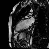 Es una prueba diagnóstica no invasiva que obtiene información morfológica y funcional del corazón y las estructuras adyacentes; de esta manera se pueden diagnosticar diferentes patologías congénitas y adquiridas, o bien realizar controles en pacientes con patología previa ya conocida. En la gran mayoría de casos es necesario el uso de contraste intravenoso (gadolinio) para completar el estudio, un tipo de contraste que raramente produce reacciones adversas. Durante la prueba, el técnico le pedirá varias veces que aguante la respiración durante 10-15 segundos para obtener las imágenes lo más claras posible. No se necesita preparación previa alguna por parte del paciente. La duración de la prueba es de aproximadamente 45-60 minutos. Está contraindicada en pacientes con marcapasos; el paciente debe avisar si es portador de implantes metálicos y/o clips quirúrgicos.
 - RM Cardíaca con estrés miocárdico
 Es una prueba diagnóstica no invasiva, que obtiene información morfológica y funcional del corazón y las estructuras adyacentes. De esta manera, se pueden diagnosticar diferentes patologías congénitas y adquiridas. Durante la prueba, el técnico le pedirá varias veces que aguante la respiración durante 10-15 segundos, para así poder obtener las imágenes lo más claras posible. En la gran mayoría de casos es necesario el uso de contraste intravenoso (gadolinio) para completar el estudio, un tipo de contraste que raramente produce reacciones adversas. Además, se introduce un fármaco (Adenosina) por vía intravenosa para ejercer sobre el corazón un efecto similar al que se produce durante el ejercicio físico. Se comparan las imágenes obtenidas en reposo y tras el "estrés" farmacológico para detectar déficits de aporte sanguíneo al ventrículo izquierdo del corazón. La duración de la prueba es de aproximadamente 45-60 minutos. Es necesario que 24 horas antes de la prueba no tome alimentos que contengan cafeína (café, té, chocolate, bebidas de cola, etc.). Está contraindicada en pacientes con marcapasos; el paciente debe avisar si es portador de implantes metálicos y/o clips quirúrgicos.
 - Angio RM Venas pulmonares
 Prueba diagnóstica no invasiva que consiste en el estudio del patrón de drenaje de las venas pulmonares mediante el empleo de un campo electromagnético y ondas de radio (con un emisor y un receptor) y de contraste paramagnético (Gadolinio). No utiliza radiación ionizante. Está indicada como en el mapa angiográfico previo en pacientes que van a ser sometidos a ablación de las venas pulmonares, así como en su seguimiento para descartar la aparición de estenosis.
 - RM Mamas
 Prueba diagnóstica no invasiva que consiste en la obtención de imágenes de alta definición anatómica de ambas mamas mediante el empleo de un campo electromagnético y ondas de radio (con un emisor y un receptor). No utiliza radiación ionizante. No requiere ninguna preparación previa. En la mayoría de casos requiere el empleo de contraste paramagnético (Gadolinio), sobre todo si la indicación es la sospecha o estudio de neoplasia mamaria. Esta prueba también está indicada en el estudio de pacientes con prótesis mamarias, ya sea en el seguimiento, para descartar complicaciones asociadas a la prótesis, o bien para descartar la sospecha de neoplasia. En el estudio de prótesis, la RM presenta una alta sensibilidad y especificidad en la detección de silicona (se aplican secuencias específicas que sólo detectan las áreas con silicona) para poder diagnosticar con la máxima fiabilidad posible la presencia de rotura tanto intra como extra-capsular.
 - RM Esternón
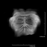 Prueba diagnóstica no invasiva que consiste en la obtención de imágenes de alta definición anatómica del esternón mediante el empleo de un campo electromagnético y ondas de radio (con un emisor y un receptor). No utiliza radiación ionizante. Está indicada para diferenciar entre lesiones benignas y malignas, en pacientes con dolor en las articulaciones esternocostales, etc. En ocasiones se deberá emplear contraste paramagnético (Gadolinio) para completar el estudio.
 - RM Axilar
 Prueba diagnóstica no invasiva que consiste en la obtención de imágenes de alta definición anatómica de la axila mediante el empleo de un campo electromagnético y ondas de radio (con un emisor y un receptor). No utiliza radiación ionizante. Está indicada en el estudio de ganglios axilares, para diferenciar entre lesiones quísticas, de contenido hemorrágico (hematomas post-quirúrgicos) y sólidas, etc. En ocasiones se deberá emplear contraste paramagnético (Gadolinio) para completar el estudio.
- Abdomen y pelvis
- RM Abdomen
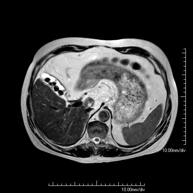 Prueba diagnóstica no invasiva que consiste en la obtención de imágenes de alta definición anatómica del abdomen mediante el empleo de un campo electromagnético y ondas de radio (con un emisor y un receptor). No utiliza radiación ionizante. En esta exploración se incluyen el hígado, páncreas, bazo, vía biliar, vesícula biliar,Entero-RM glándulas suprarrenales, riñones, aorta abdominal, vena cava inferior, estómago, duodeno,…En ocasiones se deberá emplear contraste paramagnético (Gadolinio) para caracterizar las lesiones.
 - RM Pelvis femenina
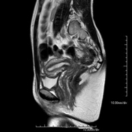 Prueba diagnóstica no invasiva que consiste en la obtención de imágenes de alta definición anatómica de la pelvis mediante el empleo de un campo electromagnético y ondas de radio (con un emisor y un receptor). No utiliza radiación ionizante. Se realiza para estudiar patologías del útero, del ovario, de las trompas y la vagina, ya sean de origen tumoral, inflamatorio o vasculares. Además permite valorar les estructuras adyacentes localizadas en la pelvis, identificando sus alteraciones. A veces requiere el uso de contraste intravenoso (Gadolinio) para caracterizar las lesiones.
 - RM Pelvis masculina
Prueba diagnóstica no invasiva que consiste en la obtención de imágenes de alta definición anatómica de la pelvis masculina mediante el empleo de un campo electromagnético y ondas de radio (con un emisor y un receptor). No utiliza radiación ionizante. No requiere de preparación previa. En algunas ocasiones requiere el empleo de contraste paramagnético (Gadolinio) para caracterizar las lesiones. Esta prueba permite valorar órganos como la vejiga urinaria, la unión entre los uréteres y la vejiga, la próstata, las vesículas seminales, la uretra, los huesos de la pelvis, etc.
- RM Hígado
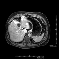 Prueba diagnóstica no invasiva que consiste en la obtención de imágenes de alta definición anatómica del hígado mediante el empleo de un campo electromagnético y ondas de radio (con un emisor y un receptor). No utiliza radiación ionizante. Se realiza para estudiar cualquier lesión localizada en el hígado, y también para valorar enfermedades inflamatorias y de depósito. Normalmente se requiere el uso de contraste paramagnético (Gadolinio) para caracterizar las lesiones. Es necesario realizar la prueba en ayunas (6 horas).
 - RM de Ronyons
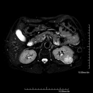 Prova diagnòstica no invasiva que consisteix en l'obtenció d'imatges d'alta definició anatòmica d'ambdós ronyons mitjançant l'ús d'un camp electromagnètic i ones de ràdio (amb un emissor i un receptor). No utilitza radiació ionitzant. Es realitza per estudiar qualsevol lesió localitzada en ambdós ronyons. Normalment es requereix l'ús de contrast paramagnètic (Gadolini) per caracteritzar les lesions.
 - RM de Glàndules Suprarenals
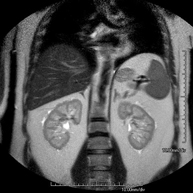 Prova diagnòstica no invasiva que consisteix en l'obtenció d'imatges d'alta definició anatòmica de les dues glàndules suprarenals mitjançant l'ús d'un camp electromagnètic i ones de ràdio (amb un emissor i un receptor). No utilitza radiació ionitzant. No necessita preparació prèvia. No requereix l'ús de contrast paramagnètic (Gadolini). Està especialment indicada en aquells pacients en els quals és imprescindible diferenciar el nòdul suprarenal benigne (l'adenoma suprarenal és el més freqüent) d'altres lesions suprarenals (tant benignes -hematomes, angiomiolipomas, etc.- com malignes).
 - RM de Melsa
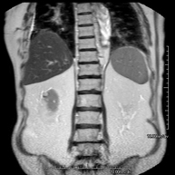 Prova diagnòstica no invasiva que consisteix en l'obtenció d'imatges d'alta definició anatòmica de la melsa mitjançant l'ús d'un camp electromagnètic i ones de ràdio (amb un emissor i un receptor). No utilitza radiació ionitzant. Es realitza per a l'estudi de qualsevol lesió dependent de la melsa. Normalment requereix l'ús de contrast paramagnètic (Gadolini) per caracteritzar les lesions.
 - Colangio-RM (RM de vesícula i via biliar)
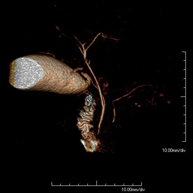 Prova diagnòstica no invasiva que consisteix en l'obtenció d'imatges d'alta definició anatòmica de la via biliar i de la vesícula biliar mitjançant l'ús d'un camp electromagnètic i ones de ràdio (amb un emissor i un receptor). No utilitza radiació ionitzant. És necessari realitzar un dejuni de 6 hores prèvies ja que, si no s'ha ingerit res, la bilis acumulada a la vesícula i a l'arbre biliar ens permetrà una millor definició anatòmica. Requereix l'obtenció de seqüències de Colangio-RM, que consisteixen en l'obtenció d'imatges de gruix molt fi (1 mm) i permet realitzar reconstruccions bidimensionals i tridimensionals sense la utilització de contrast paramagnètic (Gadolini). En algunes ocasions necessita l'ús de contrast paramagnètric (Gadolini) per caracteritzar les lesions. Està especialment indicada en pacients amb sospita de pedres a la vesícula, amb pedres a la vesícula amb sospita de desplaçament d'una de les pedres pels conductes biliars cap a l'intestí, pacients amb símptomes d'obstrucció de la via biliar, com a estudi pre-quirúrgic (mapa anatòmic de l'arbre biliar) abans de la cirurgia de la vesícula biliar, pacients amb dolor abdominal de probable origen biliar, etc.
 - Colangio-pancreàtic-RM (RM de Pàncrees, vesícula i via biliar)
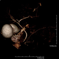 Prova diagnòstica no invasiva que consisteix en l'obtenció d'imatges d'alta definició anatòmica dels conductes de drenatge pancreàtic (principalment el conducte de Wirsung), de la via biliar i de la vesícula biliar mitjançant l'ús d'un camp electromagnètic i ones de ràdio (amb un emissor i un receptor). No utilitza radiació ionitzant. És necessari 6 hores prèvies de dejuni ja que, si no s'ha ingerit res, la bilis i els sucs pancreàtics s'acumulen i permeten una millor definició anatòmica d'aquests. En algunes ocasions es requereix l'ús de contrast paramagnètric (Gadolini) per caracteritzar les lesions. Està especialment indicada en pacients amb sospita de pedres al conducte de drenatge de la via biliar o colèdoc, de malaltia a nivell de la unió dels conductes biliars i els pancreàtics, en pacients amb pancreatitis de repetició, dolor abdominal amb probable origen biliar o pancreàtic, com a estudi anatòmic pre-quirúrgic, etc.
 - RM de Pàncrees
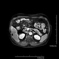 Prova diagnòstica no invasiva que consisteix en l'obtenció d'imatges d'alta definició anatòmica del pàncrees mitjançant l'ús d'un camp electromagnètic i ones de ràdio (amb un emissor i un receptor). No utilitza radiació ionitzant. Requereix dejuni de 6 hores. Permet un estudi específic pancreàtic, tant de la part glandular com dels conductes de drenatge dels sucs pancreàtics, amb una alta definició tissular. També s'estudien els vasos pancreàtics i les estructures adjacents com són la vena porta, l'artèria i la vena esplènica, el greix peri-pancreàtic, l'aorta abdominal, la vena cava inferior, etc. En la majoria dels casos s'empra contrast paramagnètic (Gadolini) per a una millor definició tissular. Aquesta prova està especialment indicada en pacients amb sospita de lesions pancreàtiques, en pacients amb neoplàsia pancreàtica coneguda com l'estudi o mapa abans de la cirurgia, en pacients amb pancreatitis crònica o aguda, etc.
 - URO-RM
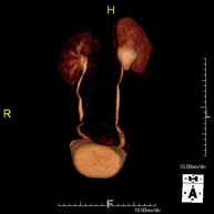 Prova diagnòstica no invasiva que consisteix en l'obtenció d'imatges d'alta definició anatòmica del sistema urinari mitjançant l'ús d'un camp electromagnètic i ones de ràdio (amb un emissor i un receptor). No utilitza radiació ionitzant. Cal utilitzar contrast paramagnètic (Gadolini) que s'expel·lirà a través del sistema urinari i ens permetrà obtenir imatges en 2D i 3D.
 - RM de Pròstata
 Prova diagnòstica no invasiva que consisteix en l'obtenció d'imatges d'alta definició anatòmica de la glàndula prostàtica mitjançant l'ús d'un camp electromagnètic i ones de ràdio (amb un emissor i un receptor). No utilitza radiació ionitzant. S'utilitza una bobina endorectal per obtenir imatges de màxima definició anatòmica de la pròstata i permetre fer l'estudi espectroscòpic (estudi a nivell molecular que ajuda a diferenciar les cèl·lules d'origen maligne o tumoral). També es realitza l'estudi amb contrast paramagnètic, que aporta una millor definició tissular. Aquesta prova dura uns 40 minuts, durant els quals el pacient haurà d'estar el més quiet possible. Requereix una neteja del còlon com a preparació prèvia. Aquesta prova està especialment indicada en aquells pacients amb sospita de neoplàsia de pròstata, amb neoplàsia de pròstata coneguda per l'estadiatge tumoral, per a l'estudi de localització del tumor prostàtic com a guia o mapa per a la biòpsia, per al seguiment dels pacients amb neoplàsia de pròstata tractats amb tractament quirúrgic o radioteràpic, davant d'una sospita de reincidència de la neoplàsia de pròstata, etc.
 - RM de Cos sencer (Total body)
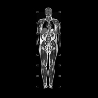 Prova diagnòstica no invasiva que consisteix en l'obtenció d'imatges d'alta definició anatòmica de tot el cos mitjançant l'ús d'un camp electromagnètic i ones de ràdio (amb un emissor i un receptor). No utilitza radiació ionitzant. És una prova molt important en la recerca de metàstasi en pacients amb neoplàsia coneguda. No requereix preparació prèvia. No és necessari l'ús de contrast paramagnètic (Gadolini).
 - Angio-RM d'Aorta abdominal
 Prova diagnòstica no invasiva que consisteix en l'obtenció d'imatges d'alta definició anatòmica de tot el cos mitjançant l'ús d'un camp electromagnètic i ones de ràdio (amb un emissor i un receptor). No utilitza radiació ionitzant. És una prova molt important en la recerca de metàstasi en pacients amb neoplàsia coneguda. No requereix preparació prèvia. No és necessari l'ús de contrast paramagnètic (Gadolini).
 - Angio RM d'Aorta-ilíaca
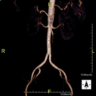 Prova diagnòstica no invasiva que consisteix en l'estudi de l'artèria aorta abdominal i de les artèries ilíaques, amb l'obtenció d'imatges d'alta definició anatòmica mitjançant l'ús d'un camp electromagnètic i ones de ràdio (amb un emissor i un receptor). És indispensable l'ús de contrast paramagnètic (Gadolini). No utilitza radiació ionitzant. La qualitat de les imatges permet realitzar reconstruccions en 2D i 3D. Aquesta prova està especialment indicada com a estudi prequirúrgic (mapa vascular) abans d'intervencions percutànies o quirúrgiques d'aorta abdominal i artèries ilíaques, per a l'estudi complementari en pacients amb isquèmia de membres inferiors, etc.
 - Angio RM Arterial d'extremitats inferiors
 Prova diagnòstica no invasiva que consisteix en l'estudi vascular del sector aorto-ilíac i dels vasos arterials de les dues extremitats inferiors, am l'obtenció d'imatges d'alta definició anatòmica mitjançant l'ús d'un camp electromagnètic i ones de ràdio (amb un emissor i un receptor). És indispensable l'ús de contrast paramagnètic (Gadolini). No obstant, no utilitza radiació ionitzant. La qualitat de les imatges permet realitzar reconstruccions en 2D i 3D. Està especialment indicat en aquells pacients en els que hi ha sospita de malaltia vascular a les dues extremitats, en pacients amb malaltia vascular de les dues extremitats com a mapa vascular abans del tractament (percutani o quirúrgic), com a mapa vascular prequirúrgic en pacients amb lesions òssies o musculars que requereixin cirurgia, etc.
 - Angio-RM d'Artèries renals
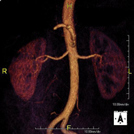 Prova diagnòstica no invasiva que consisteix en l'estudi de les artèries renals per obtenir imatges d'alta definició anatòmica mitjançant l'ús d'un camp electromagnètic i ones de ràdio (amb un emissor i un receptor). És indispensable l'ús de contrast paramagnètic (Gadolini). No obstant, no utilitza radiació ionitzant. La qualitat de les imatges permet realitzar reconstruccions en 2D i 3D. Aquesta prova està indicada en aquells pacients que pateixen d'HTA refractària al tractament, en pacients amb lesions renals per tenir un mapa "vascular" pre-quirúrgic, etc.
 - Sencer-RM
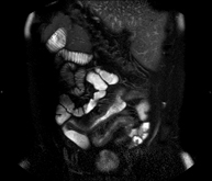 Prova diagnòstica no invasiva que consisteix per obtenir d'imatges d'alta definició anatòmica de l'intestí mitjançant l'ús d'un camp electromagnètic i ones de ràdio (amb un emissor i un receptor). No utilitza radiació ionitzant. Normalment requereix l'ús de contrast paramagnètic (Gadolini). Està indicada principalment en el diagnòstic, el seguiment i el control de la resposta al tractament dels pacients amb Malaltia de Crohn.
 - RM Rectal
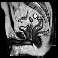 Prova diagnòstica no invasiva que consisteix en l'obtenció d'imatges d'alta definició anatòmica del recte mitjançant l'ús d'un camp electromagnètic i ones de ràdio (amb un emissor i un receptor). No utilitza radiació ionitzant. Normalment no requereix l'ús de contrast paramagnètic (Gadolini). Està indicada principalment en el diagnòstic, l'estadiatge i el seguiment del Càncer de recte.
 - RM de Paret abdominal
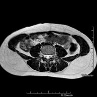 Prova diagnòstica no invasiva que consisteix en l'obtenció d'imatges d'alta definició anatòmica de la paret abdominal mitjançant l'ús d'un camp electromagnètic i ones de ràdio (amb un emissor i un receptor). No utilitza radiació ionitzant. Està indicada en l'estudi de lesions de la paret abdominal i solen correspondre a bonys que detecta el pacient o a dolors en aquesta regió anatòmica. De vegades s'ha d'utilitzar contrast paragmanètic (Gadolini) per completar l'estudi.
 - RM Escrotal (testicular)
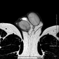 Prova diagnòstica no invasiva que consisteix en l'obtenció d'imatges d'alta definició anatòmica de la regió escrotal mitjançant l'ús d'un camp electromagnètic i ones de ràdio (amb un emissor i un receptor). No utilitza radiació ionitzant. De vegades s'ha d'emprar contrast paragmanètic (Gadolini) per completar l'estudi. S'utilitza per a l'estudi detallat dels testicles, per identificar possibles tumors i diverses patologies i per visualitzar alteracions de les estructures adjacents.
 - RM peniana
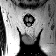 Prova diagnòstica no invasiva que consisteix en l'obtenció d'imatges d'alta definició anatòmica de la regió peniana mitjançant l'ús d'un camp electromagnètic i ones de ràdio (amb un emissor i un receptor). No utilitza radiació ionitzant. De vegades s'ha d'emprar contrast paragmanètic (Gadolini) per completar l'estudi. Normalment es realitza per estudiar la patologia peniana d'origen traumàtic i per visualitzar alteracions de les estructures adjacents.
- Múscul – esquelètic
- RM d'Espatlla
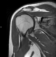 Exploració per a l'estudi de lesions en els tendons, els músculs i les articulacions. La seva principal utilitat resideix en diagnosticar les lesions dels tendons del braçal rotatori. Té una durada aproximada de 20 minuts. No utilitza radiació ionitzant.
 - RM de Braç
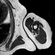 Exploració per a l'estudi de lesions en els tendons, els músculs i els nervis perifèrics. També és útil en l'estudi de tumors. La durada aproximada és de 20 minuts. No utilitza radiació ionitzant.
 - RM de Colze
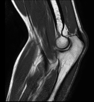 Exploració per a l'estudi de lesions en els tendons, els músculs i les articulacions. Molt útil per valorar trencaments del tendó del bíceps i la epicondilitis (colze de tenista). La durada aproximada és de 20 minuts. No utilitza radiació ionitzant.
 - RM Antebrazo
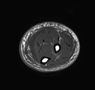 Exploración para el estudio de lesiones en tendones, músculos y nervios periféricos. Tiene una duración aproximada de 18 minutos. No emplea radiación ionizante.
 - RM Muñeca / carpo
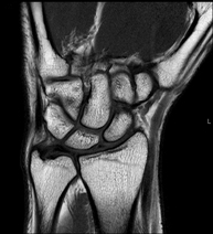 Exploración para el estudio de lesiones en tendones músculos y articulación. Muy útil para la valoración de pequeñas fracturas inadvertidas, lesiones de ligamentos, y procesos inflamatorios y degenerativos (artritis y artrosis). Tiene una duración aproximada de 20 minutos. No emplea radiación ionizante.
 - RM Mano / dedos
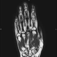 Exploración para el estudio de lesiones en tendones, ligamentos y pequeñas articulaciones. Es la prueba que mejor permite diagnosticar las frecuentes capsulitis por traumatismo, la artrosis y las roturas de tendones. Tiene una duración aproximada de 20 minutos. No emplea radiación ionizante.
 - Hip MRI
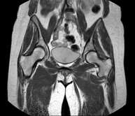
Examination for the study of injuries to tendons, muscles and hip joints. Enables early detection of hip osteoarthritis. It is very useful for detecting bursitis and dynamic osteopathy of the pubis, which is common in athletes. It lasts approximately 20 minutes. It is a radiation-free procedure.
- Sacroiliac MRI
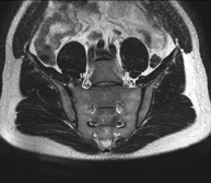
Study specifically designed to assess these joints and their inflammation in patients suffering from ankylosing spondylitis. It is also useful in patients with trauma and possible fractures of the sacrum and coccyx. It lasts approximately 16 minutes. It is a radiation-free procedure.
- Gluteal MRI
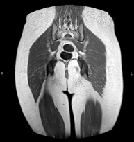
Examination to study the muscles and tendons that originate at this level, such as the hamstring tendons, which are frequently injured in athletes. It lasts approximately 16 minutes. It is a radiation-free procedure.
- Thigh MRI
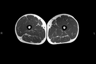
Ideal examination for studying injuries to the hamstrings and quadriceps, which are frequently injured in athletes. It also allows for a good assessment of tendons and peripheral nerves. It lasts approximately 20 minutes. It is a radiation-free procedure.
- Knee MRI
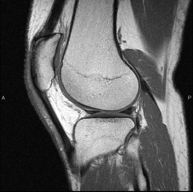
Examination to study joint injuries, such as meniscal tears and cruciate ligament tears (only detectable through this test), chondropathy or cartilage wear, and a multitude of other disorders resulting from sports activities and degenerative changes (osteoarthritis). It lasts approximately 18 minutes. It is a radiation-free procedure.
- Leg MRI
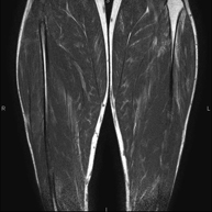
Examination to study injuries to tendons, muscles and peripheral nerves. Very useful for diagnosing fibrillar tears in twins. It lasts approximately 18 minutes. It is a radiation-free procedure.
- Ankle MRI
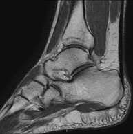
Examination to study the joint and its frequent ligament injuries (sprains) as well as damage to other structures such as cartilage or bone. It is also very useful for diagnosing disorders of the Achilles tendon (tendinitis, ruptures). Duration: approximately 20 minutes. It is a radiation-free procedure.
- Foot MRI
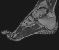
Examination for the study of injuries to tendons, muscles and small joints. Allows for effective assessment of cartilage injuries. It is also very useful for completing the preliminary ultrasound examination. It is a radiation-free procedure. It lasts approximately 20 minutes.
- Shoulder MRI arthrography
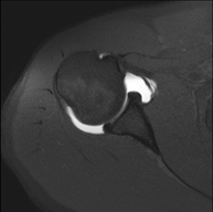
Examination to study injuries to small anatomical structures of the joint that are commonly injured in patients suffering from dislocation or chronic instability. Prior to the examination, a contrast fluid is injected into the joint, guided by X-ray imaging. The total duration of the two procedures is 50 minutes.
- Wrist MRI arthrography
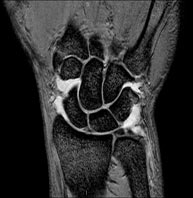
Examination to study injuries to small anatomical structures in joints, such as ligaments and cartilage. Prior to the examination, a contrast fluid is injected into a joint, guided by X-ray imaging. The total duration of the two procedures is 50 minutes.
- Artro-RM de Maluc
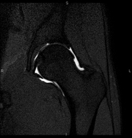 Exploració per estudiar les lesions en petites estructures anatòmiques de l'articulació que solen produir-se en pacients que pateixen luxació o pinçament (disminució de l'espai). L'estudi ve precedit per una injecció de contrast a l'interior de l'articulació, realitzada sota control de raigs X. La durada total dels dos procediments és de 50 minuts.
 - Artro-RM de Genoll
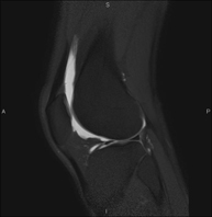 Exploració per a l'estudi de les lesions en petites estructures anatòmiques de l'articulació, com ara el cartílag i els meniscs, principalment els ja operats. L'estudi ve precedit per una injecció de contrast a l'interior de l'articulació, realitzada sota control de raigs X. La durada total dels dos procediments és de 50 minuts.
 - Artro-RM de Turmell
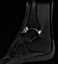 Exploració per a l'estudi de les lesions en petites estructures anatòmiques de l'articulació que solen produir-se en pacients que pateixen luxació o inestabilitat crònica. L'estudi ve precedit per una injecció de contrast a l'interior de l'articulació, realitzada sota control de raigs X. La durada total dels dos procediments és de 50 minuts.
- Columna
- RM Columna Cervical
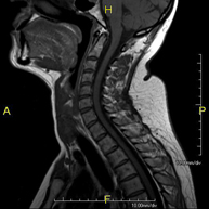 Prova diagnòstica no invasiva que consisteix en l'obtenció d'imatges d'alta definició anatòmica de la columna cervical, mitjançant l'ús d'un camp electromagnètic i ones de ràdio (amb un emissor i un receptor). No utilitza radiació ionitzant. Indicacions: traumatisme, degeneració de la columna i hèrnies.
 - RM Columna Dorsal
 Prova diagnòstica no invasiva que consisteix en l'obtenció d'imatges d'alta definició anatòmica de la columna dorsal, mitjançant l'ús d'un camp electromagnètic i ones de ràdio (amb un emissor i un receptor). No utilitza radiació ionitzant. Indicacions: traumatisme, problemes degeneratius, hèrnies i tumors.
 - RM Columna Lumbar
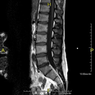 Prova diagnòstica no invasiva que consisteix en l'obtenció d'imatges d'alta definició anatòmica de la columna lumbar i sacre mitjançant l'ús d'un camp electromagnètic i ones de ràdio (amb un emissor i un receptor). No utilitza radiació ionitzant. Indicacions: traumatismes, ciàtica, hèrnies discals, tumors i infeccions.
 - RM de Sacre-còccix
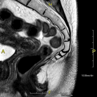 Prova diagnòstica no invasiva que consisteix en l'obtenció d'imatges d'alta definició anatòmica del sacre i còccix mitjançant l'ús d'un camp electromagnètic i ones de ràdio (amb un emissor i un receptor). No utilitza radiació ionitzant. Indicacions: dolor sacrococcigi i traumatisme.
 - RM Columna cervical + dorsal
Prova diagnòstica no invasiva que consisteix en l'obtenció d'imatges d'alta definició anatòmica de la columna cervical i dorsal mitjançant l'ús d'un camp electromagnètic i ones de ràdio (amb un emissor i un receptor). No utilitza radiació ionitzant. Indicacions: traumatisme, degeneració de la columna i hèrnies.
- RM Columna dorsal + lumbar
Prova diagnòstica no invasiva que consisteix en l'obtenció d'imatges d'alta definició anatòmica de la columna dorsal i lumbar, mitjançant l'ús d'un camp electromagnètic i ones de ràdio (amb un emissor i un receptor). No utilitza radiació ionitzant. Indicacions: traumatisme, degeneració de la columna i hèrnies.
- RM Columna completa
Prova diagnòstica no invasiva que consisteix en l'obtenció d'imatges d'alta definició anatòmica de la columna cervical, dorsal i lumbar, mitjançant l'ús d'un camp electromagnètic i ones de ràdio (amb un emissor i un receptor). No utilitza radiació ionitzant. Indicacions: detecció de metàstasi, escoliosi i traumatisme.
- Pediatría
- RM Cráneo
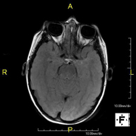 Prueba diagnóstica no invasiva que consiste en la obtención de imágenes de alta definición anatómica del cráneo mediante el empleo de un campo electromagnético y ondas de radio (con un emisor y un receptor). No utiliza radiación ionizante. En ocasiones se deberá emplear contraste paramagnético (Gadolinio) para completar el estudio.
 - RM Cuello
Prueba diagnóstica no invasiva que consiste en la obtención de imágenes de alta definición anatómica del cuello mediante el empleo de un campo electromagnético y ondas de radio (con un emisor y un receptor). No utiliza radiación ionizante. En ocasiones se deberá emplear contraste paramagnético (Gadolinio) para completar el estudio.
- RM Tórax
 Prueba diagnóstica no invasiva que consiste en la obtención de imágenes de alta definición anatómica del tórax mediante el empleo de un campo electromagnético y ondas de radio (con un emisor y un receptor). No utiliza radiación ionizante. Está indicada en aquellas lesiones pulmonares en las que debe descartarse si existe infiltración del mediastino o de la pared torácica, para diferenciar si una lesión torácica es sólida o quística, etc. En ocasiones se deberá emplear contraste paramagnético (Gadolinio) para completar el estudio.
 - RM Abdomen
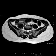 Prueba diagnóstica no invasiva que consiste en la obtención de imágenes de alta definición anatómica del abdomen mediante el empleo de un campo electromagnético y ondas de radio (con un emisor y un receptor). No utiliza radiación ionizante. En esta exploración se incluyen el hígado, páncreas, bazo, vía biliar, vesícula biliar, glándulas suprarrenales, riñones, aorta abdominal, vena cava inferior, estómago, duodeno, etc. En ocasiones se deberá emplear contraste paramagnético (Gadolinio) para caracterizar las lesiones.
 - URO-RM
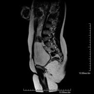 Prueba diagnóstica no invasiva que consiste en la obtención de imágenes de alta definición anatómica del sistema urinario mediante el empleo de un campo electromagnético y ondas de radio (con un emisor y un receptor). No utiliza radiación ionizante. Requiere el uso de contraste paramagnético (Gadolinio) que se excretará por el sistema urinario y nos permitirá obtener imágenes en 2D y 3D.
 - RM Bajo sedación
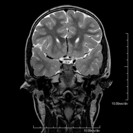 Prueba diagnóstica no invasiva que consiste en la obtención de imágenes de alta definición anatómica de cualquier región del cuerpo mediante el empleo de un campo electromagnético y ondas de radio (con un emisor y un receptor). No utiliza radiación ionizante. Se realiza bajo sedación con la colaboración del equipo de Anestesia. En los pacientes pediátricos es de gran ayuda porque permite realizar exploraciones sin artefactos de movimiento (lactantes, niños de corta edad, etc. en los que la situación clínica lo requiera).
 - RM Fetal
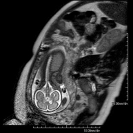 Prueba diagnóstica no invasiva que consiste en la obtención de imágenes de alta definición anatómica del feto en la mujer gestante mediante el empleo de un campo electromagnético y ondas de radio (con un emisor y un receptor). No utiliza radiación ionizante. Se puede realizar a partir de la semana 12 de gestación, siendo segura tanto para el feto como para la madre. Se utiliza en el caso de sospechar alguna anomalía congénita morfológica.
 - RM Muscular
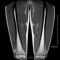 Prueba diagnóstica no invasiva que consiste en la obtención de imágenes de alta definición anatómica de la musculatura a estudio mediante el empleo de un campo electromagnético y ondas de radio (con un emisor y un receptor). No utiliza radiación ionizante. En escasas ocasiones se requiere el uso de contraste paramagnético (Gadolinio) para mejor definición de las lesiones.
 - RM Huesos y articulaciones
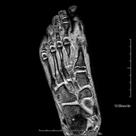 Prueba diagnóstica no invasiva que consiste en la obtención de imágenes de alta definición anatómica de huesos y articulaciones mediante el empleo de un campo electromagnético y ondas de radio (con un emisor y un receptor). No utiliza radiación ionizante. En escasas ocasiones se requiere el uso de contraste paramagnético (Gadolinio) para mejor definición de las lesiones.
- Mama
- RM Mamas
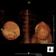 Prueba diagnóstica no invasiva que consiste en la obtención de imágenes de alta definición anatómica de ambas mamas mediante el empleo de un campo electromagnético y ondas de radio (con un emisor y un receptor). No utiliza radiación ionizante. No requiere ninguna preparación previa. En la mayoría de casos requiere el empleo de contraste paramagnético (Gadolinio), sobre todo si la indicación es la sospecha o estudio de neoplasia mamaria. Esta prueba también está indicada en el estudio de pacientes con prótesis mamarias, ya sea en el seguimiento, para descartar complicaciones asociadas a la prótesis, o bien, para descartar la sospecha de neoplasia. En el estudio de prótesis la RM presenta una alta sensibilidad y especificidad en la detección de silicona (se aplican secuencias específicas que sólo detectan las áreas con silicona) parar poder diagnosticar con la máxima fiabilidad posible la presencia de roturas tanto intra como extracapsulares.
- Corazón
- RM Cardíaca
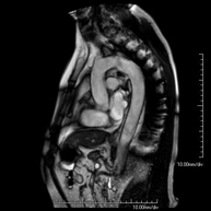 Es una prueba diagnóstica no invasiva, que obtiene información morfológica y funcional del corazón y las estructuras adyacentes. De esta manera, se pueden diagnosticar diferentes patologías congénitas y adquiridas, o bien realizar controles en pacientes con patología previa ya conocida. En la gran mayoría de casos es necesario el uso de contraste intravenoso (gadolinio) para completar el estudio, un tipo de contraste que raramente produce reacciones adversas. Durante la prueba, el técnico le pedirá varias veces que aguante la respiración durante 10-15 segundos, para obtener las imágenes lo más claras posible. No se necesita preparación previa alguna por parte del paciente. La duración de la prueba es de aproximadamente 45 - 60 minutos. Está contraindicada en pacientes con marcapasos, y el paciente debe avisar si es portador de implantes metálicos y/o clips quirúrgicos.
 - RM Cardíaca con estrés miocárdico
 Es una prueba diagnóstica no invasiva, que obtiene información morfológica y funcional del corazón y las estructuras adyacentes. De esta manera, se pueden diagnosticar diferentes patologías congénitas y adquiridas. Durante la prueba, el técnico le pedirá varias veces que aguante la respiración durante 10-15 segundos, para así poder obtener las imágenes lo más claras posible. En la gran mayoría de casos es necesario el uso de contraste intravenoso (gadolinio) para completar el estudio, un tipo de contraste que raramente produce reacciones adversas. Además, se introduce un fármaco (Adenosina) por vía intravenosa, para ejercer sobre el corazón un efecto similar al que se produce durante el ejercicio físico. Se comparan las imágenes obtenidas en reposo y tras el "estrés" farmacológico para detectar déficits de aporte sanguíneo al ventrículo izquierdo del corazón. La duración de la prueba es de aproximadamente 45 - 60 minutos. Es necesario que 24 horas antes de la prueba no tome alimentos que contengan cafeína (café, té, chocolate, bebidas de cola…). Está contraindicada en pacientes con marcapasos, y el paciente debe avisar si es portador de implantes metálicos y/o clips quirúrgicos.
 - Angio-RM Venas pulmonares
 Prueba diagnóstica no invasiva que consiste en el estudio del patrón de drenaje de las venas pulmonares mediante el empleo de un campo electromagnético y ondas de radio (con un emisor y un receptor) y de contraste paramagnético (Gadolinio). No utiliza radiación ionizante. Está indicada como mapa angiográfico previo en pacientes que van a ser sometidos a ablación de las venas pulmonares, así como en su seguimiento para descartar la aparición de estenosis.
- Estudios vasculares
- Angio-RM Intracraneal
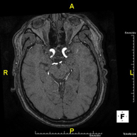 Prueba diagnóstica no invasiva que consiste en la obtención de imágenes de alta definición anatómica de las arterias cerebrales, mediante el empleo de un campo electromagnético y ondas de radio (con un emisor y un receptor). No utiliza radiación ionizante. En la mayoría de los casos es necesario el empleo de contraste paramagnético (Gadolinio). Permite un estudio angiográfico no invasivo gracias a la inyección de Gadolinio con posterior reconstrucción en 2D y 3D, gracias a estaciones de trabajo especializadas. Indicaciones: Malformaciones vasculares, aneurismas de arterias cerebrales, arteriosclerosis.
 - Angio-RM Troncos supraaórticos
Prueba diagnóstica no invasiva que consiste en la obtención de imágenes de alta definición anatómica de las arterias carótidas y vertebrales a nivel de cuello, mediante el empleo de un campo electromagnético y ondas de radio (con un emisor y un receptor). No utiliza radiación ionizante. En la mayoría de los casos es necesario el empleo de contraste paramagnético (Gadolinio). Permite un estudio angiográfico no invasivo gracias a la inyección de Gadolinio con posterior reconstrucción en 2D y 3D, gracias a estaciones de trabajo especializadas. Indicaciones: Problemas circulatorios cerebrales, síncope.
- Angio-RM Aorta Torácica
 Prueba diagnóstica no invasiva que consiste en la obtención de imágenes de alta definición anatómica de la aorta torácica (principal arteria del tórax) mediante el empleo de un campo electromagnético y ondas de radio (con un emisor y un receptor). No utiliza radiación ionizante. En la mayoría de los casos es necesario el empleo de contraste paramagnético (Gadolinio). Permite un estudio estudio angiográfico no invasivo gracias a la inyección de Gadolinio con posterior reconstrucción en 2D y 3D, gracias a estaciones de trabajo especializadas. También incluye es estudio de la válvula aórtica, información impresecindible en el caso que el paciente necesite cirugía. Esta prueba está especialmente indicada en pacientes que requieren tratamiento quirúrgico ( como mapa vascular pre-quirúrgico), en el seguimiento de pacientes con aneurismas de aorta, etc.
 - Angio-RM Aorta Abdominal
 Prueba diagnóstica no invasiva que consiste en el estudio de la arteria aorta abdominal, obteniendo imágenes de alta definición anatómica mediante el empleo de un campo electromagnético y ondas de radio (con un emisor y un receptor). Es indispensable el uso de contraste intravenoso paramagnético (gadolinio). Sin embargo, no utiliza radiación ionizante. La calidad de las imágenes permite realizar reconstrucciones en 2D y 3D. Está indicado en aquellos pacientes con enfermedad vascular (aterosclerosis), estudio de aneurismas, en estudios pre-quirúrgicos de lesiones adyacentes a la aorta abdominal como "mapa" vascular…
 - Angio-RM Aorta iliaca
 Prueba diagnóstica no invasiva que consiste en el estudio de la arteria aorta abdominal, obteniendo imágenes de alta definición anatómica mediante el empleo de un campo electromagnético y ondas de radio (con un emisor y un receptor). Es indispensable el uso de contraste intravenoso paramagnético (gadolinio). Sin embargo, no utiliza radiación ionizante. La calidad de las imágenes permite realizar reconstrucciones en 2D y 3D. Esta prueba está especialmente indicada como estudio pre-quirúrgico (mapa vascular) antes de intervenciones percutáneas o quirúrgicas de aorta abdominal, estudio complementario en pacientes con isquemia de miembros inferiores…
 - Angio-RM Arterias renales
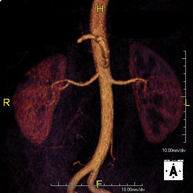 Prueba diagnóstica no invasiva que consiste en el estudio de la arteria aorta abdominal, obteniendo imágenes de alta definición anatómica mediante el empleo de un campo electromagnético y ondas de radio (con un emisor y un receptor). Es indispensable el uso de contraste intravenoso paramagnético (gadolinio). Sin embargo, no utiliza radiación ionizante. La calidad de las imágenes permite realizar reconstrucciones en 2D y 3D. Esta prueba está indicada por ejemplo en aquellos pacientes que sufren de HTA refractaria al tratamiento, en pacientes con lesiones renales para tener un mapa "vascular" pre-quirúrgico…
 - Angio-RM Arterial extremidades inferiores
 Prueba diagnóstica no invasiva que consiste en el estudio de la arteria aorta abdominal, obteniendo imágenes de alta definición anatómica mediante el empleo de un campo electromagnético y ondas de radio (con un emisor y un receptor). Es indispensable el uso de contraste intravenoso paramagnético (gadolinio). Sin embargo, no utiliza radiación ionizante. La calidad de las imágenes permite realizar reconstrucciones en 2D y 3D. Está especialmente indicado en aquellos pacientes en los que hay sospecha de enfermedad vascular de ambas extremidades, en pacientes con enfermedad vascular de ambas extremidades como mapa vascular antes del tratamiento (percutáneo o quirúrgico), como mapa vascular pre-quirúrgico en pacientes con lesiones óseas o musculares que requieran cirugía…
 - Angio-RM Lesión partes blandas
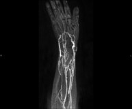 Prueba diagnóstica no invasiva que consiste en el estudio de la vascularización de cualquier lesión de partes blandas (piel, músculo, tendón,etc.) obteniendo imágenes de alta definición anatómica mediante el empleo de un campo electromagnético y ondas de radio (con un emisor y un receptor). Es indispensable el uso de contraste intravenoso paramagnético (gadolinio). Sin embargo, no utiliza radiación ionizante. La calidad de las imágenes permite realizar reconstrucciones en 2D y 3D. Está especialmente indicado como mapa vascular pre-quirúrgico en pacientes con lesiones musculares, cutáneas, subcutáneas, etc.
 - Angio-RM venas pulmonares
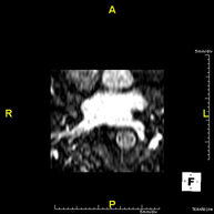 Prueba diagnóstica no invasiva que consiste en el estudio del patrón de drenaje de las venas pulmonares mediante el empleo de un campo electromagnético y ondas de radio (con un emisor y un receptor) y de contraste paramagnético (Gadolinio). No utiliza radiación ionizante. Está indicada como mapa angiográfico previo en pacientes que van a ser sometidos a ablación de las venas pulmonares, así como en su seguimiento para descartar la aparición de estenosis.
 - Angio-RM extremidades superiores
 Prueba diagnóstica no invasiva que consiste en el estudio de las arterias de la cintura escapular, brazo, antebrazo y mano obteniendo imágenes de alta definición anatómica mediante el empleo de un campo electromagnético y ondas de radio (con un emisor y un receptor). Es indispensable el uso de contraste intravenoso paramagnético (gadolinio). Sin embargo, no utiliza radiación ionizante. La calidad de las imágenes permite realizar reconstrucciones en 2D y 3D. Está especialmente indicado en aquellos pacientes en los que hay sospecha de enfermedad vascular de ambas extremidades, en pacientes con enfermedad vascular de ambas extremidades como mapa vascular antes del tratamiento (percutáneo o quirúrgico), como mapa vascular pre-quirúrgico en pacientes con lesiones óseas o musculares que requieran cirugía…